Introduction
Introduction
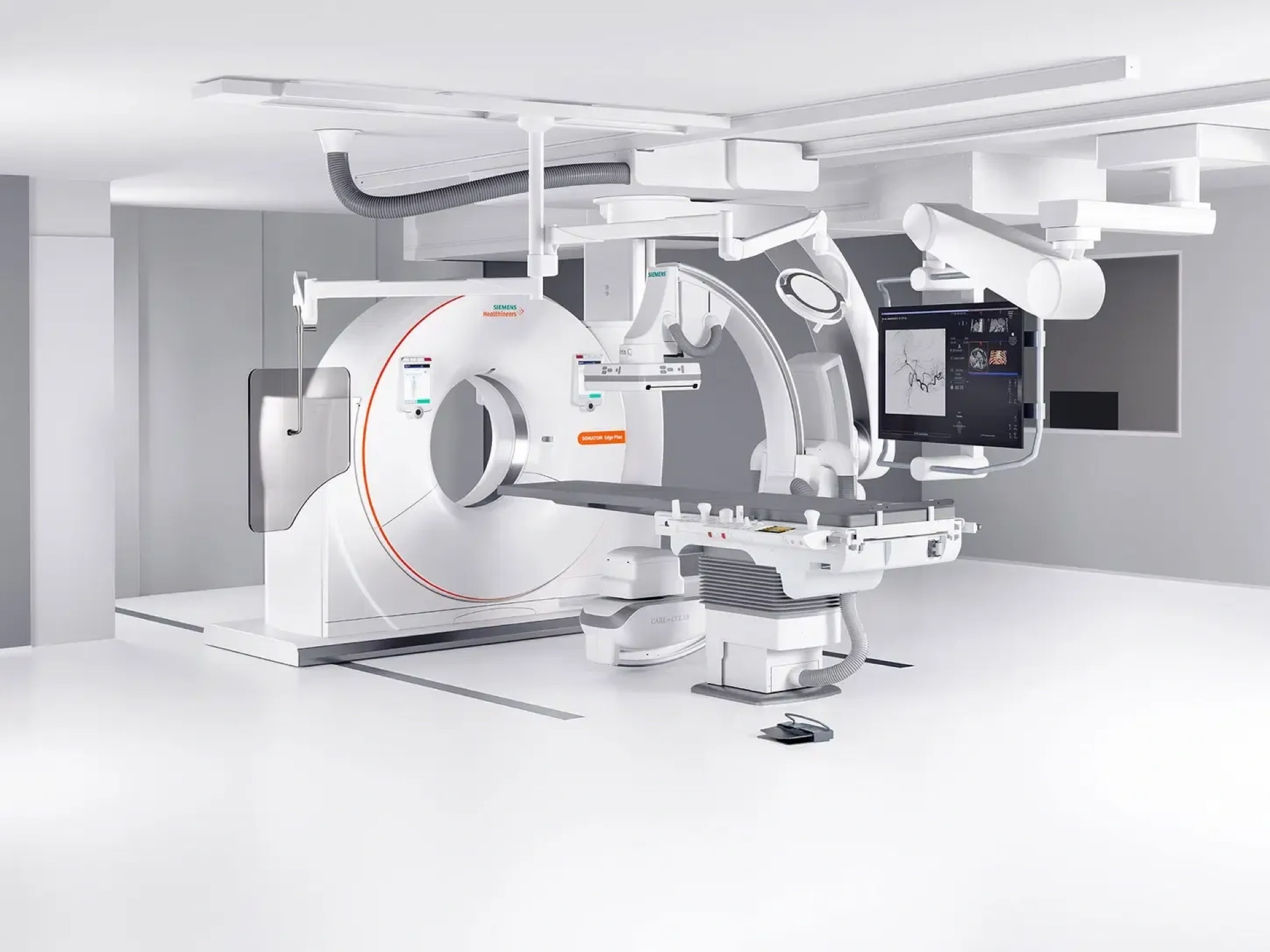
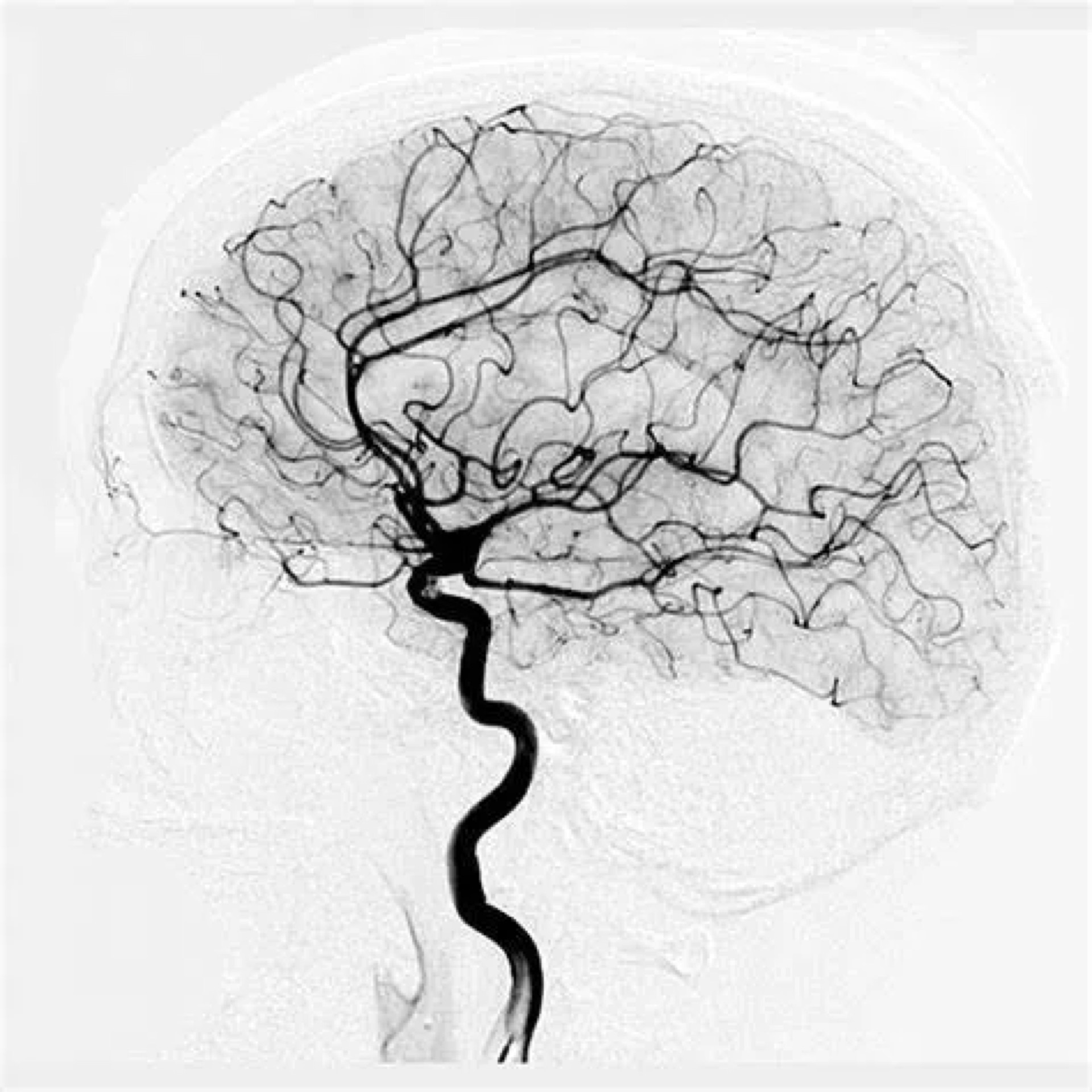
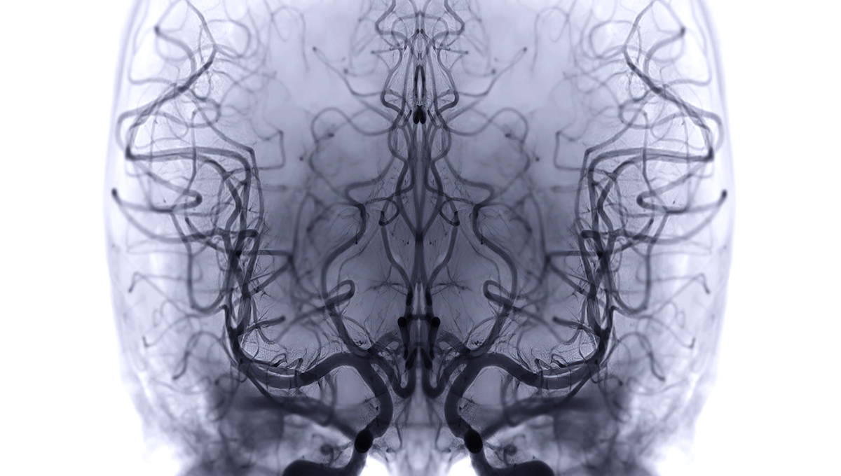
Cerebral angiography is a minimally invasive diagnostic procedure that uses contrast dye and X-ray imaging to produce detailed pictures of the blood vessels in the brain and neck. It remains the gold standard for evaluating vascular conditions of the brain.
This advanced imaging technique allows neurovascular specialists to visualize the intricate network of arteries and veins, identifying abnormalities with a level of precision that non-invasive imaging cannot match.