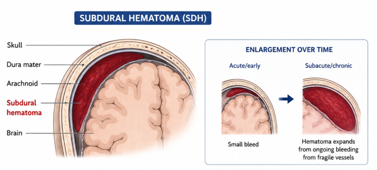
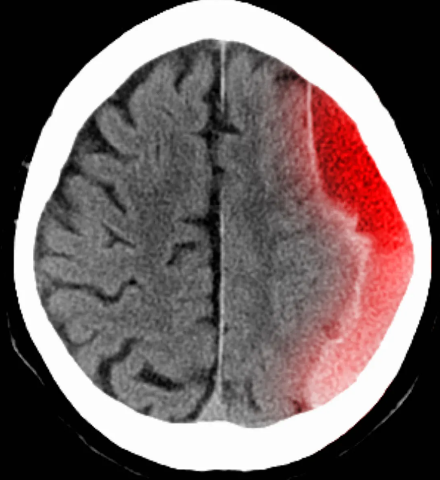
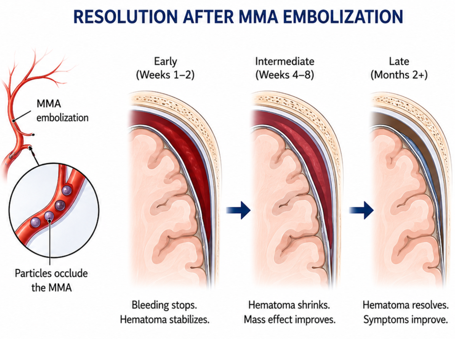
A subdural hematoma is a collection of blood that forms between the brain and the dura mater — the tough outer membrane that covers the brain. Chronic subdural hematomas develop slowly over days to weeks, often after a minor head injury or even without any remembered trauma, and are particularly common in older adults.
Symptoms can include headaches, confusion, weakness on one side of the body, difficulty walking, and changes in personality or alertness. Because these symptoms may develop gradually and mimic other conditions, subdural hematomas can sometimes go undiagnosed without proper imaging.

Acute Subdural Hematoma — Emergency
An acute subdural hematoma develops rapidly after a significant head injury and is a medical emergency. Seek emergency care immediately for:
Chronic Subdural Hematoma — Needs Evaluation Within Days
Chronic subdural hematomas develop slowly and symptoms may be subtle. Contact your doctor promptly if you experience:

The standard treatment for subdural hematomas has been surgical drainage — either through a burr hole (small opening in the skull) or a larger craniotomy. While effective in many cases, surgery carries risks including infection, bleeding, and recurrence. Some patients, particularly elderly individuals or those with significant medical conditions, may not be ideal surgical candidates.
Embolization is a newer, minimally invasive option that seals the blood vessels supplying the outer membrane of the hematoma — specifically the middle meningeal artery. By blocking these vessels, the bleeding stops and the hematoma can resolve without open surgery. This approach is performed through a small catheter, requires no incision in the skull, and dramatically reduces recovery time and risk.

This minimally invasive procedure is performed in an angiography suite and typically takes less than an hour.

